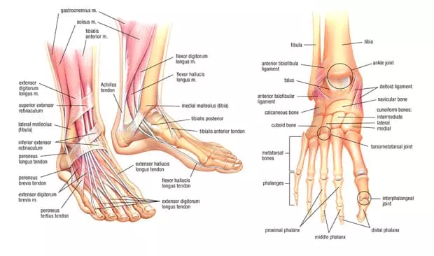
Foot Anatomy
The human foot combines mechanical complexity and structural strength. The ankle serves as foundation, shock absorber, and propulsion engine. The foot can sustain enormous pressure (several tons over the course of a one-mile run) and provides flexibility and resiliency.

The foot and ankle contain:
- • 26 bones (One-quarter of the bones in the human body are in the feet.)
- • 33 joints
- • More than 100 muscles, tendons and ligaments (Tendons are fibrous tissues that connect muscles to bones and ligaments are fibrous tissues that connect bones to other bones.) and
- • A network of blood vessels, nerves, skin, and soft tissue.
These components work together to provide the body with support, balance, and mobility. A structural flaw or malfunction in any one part can result in the development of problems elsewhere in the body. Abnormalities in other parts of the body can lead to problems in the feet.
The forefoot is composed of the five toes (called phalanges) and their connecting long bones (metatarsals). Each toe (phalanx) is made up of several small bones. The big toe (also known as the hallux) has two phalanx bones—distal and proximal. It has one joint, called the interphalangeal joint. The big toe articulates with the head of the first metatarsal and is called the first metatarsophalangeal joint (MTPJ for short). Underneath the first metatarsal head are two tiny, round bones called sesamoids. The other four toes each have three bones and two joints. The phalanges are connected to the metatarsals by five metatarsal phalangeal joints at the ball of the foot. The forefoot bears half the body’s weight and balances pressure on the ball of the foot.
The midfoot has five irregularly shaped tarsal bones, forms the foot’s arch, and serves as a shock absorber. The bones of the midfoot are connected to the forefoot and the hindfoot by muscles and the plantar fascia (arch ligament).
The hindfoot is composed of three joints and links the midfoot to the ankle (talus). The top of the talus is connected to the two long bones of the lower leg (tibia and fibula), forming a hinge that allows the foot to move up and down. The heel bone (calcaneus) is the largest bone in the foot. It joins the talus to form the subtalar joint. The bottom of the heel bone is cushioned by a layer of fat.
